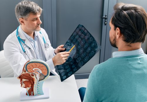
Nerve damage can cause a wide range of symptoms, from pain and numbness to muscle weakness and loss of function, significantly impacting quality of life. The central nervous system, comprising the brain and spinal cord, works alongside the peripheral nervous system to ensure overall body function and posture. Diagnosing nerve injuries, whether due to trauma, compression, or underlying medical conditions, often requires detailed imaging to visualize the affected structures. Among the advanced diagnostic tools available, Magnetic Resonance Imaging (MRI) has emerged as a critical technology for identifying and assessing nerve damage.
In this blog, we’ll explore how MRI detects nerve damage, its advantages and limitations, and invite you to visit our Atlanta medical clinic for expert evaluation and advanced imaging solutions.
Understanding MRI and Nerve Damage
Magnetic resonance imaging (MRI) is a powerful tool that plays a crucial role in diagnosing nerve damage. It uses strong magnetic fields and radio waves to create detailed images of soft tissues, including nerves. This non-invasive imaging technique is especially valuable in visualizing the peripheral nervous system, which includes all the nerves outside the brain and spinal cord. MRI is a key component of peripheral nerve imaging, providing advanced diagnostic insights into nerve injuries.
Overview of MRI Technology and Its Role in Visualizing Soft Tissues
MRI technology excels in providing high-resolution images of soft tissues. Unlike X-rays or CT scans, which primarily show bone structures, MRI can differentiate between various tissue types. Peripheral nerve MRI is a specialized imaging technique that offers high-resolution images of peripheral nerves, essential for identifying abnormalities in nerves, muscles, and surrounding soft tissues.
When a patient undergoes an MRI scan, the machine generates detailed cross-sectional images. These images can reveal important information about the condition of peripheral nerves, helping healthcare providers assess any potential damage or pathology.
Importance of MRI in Diagnosing Peripheral Nerve Injury
MRI is particularly significant when used to diagnose peripheral nerve injury. Conditions such as nerve compression, trauma, or inflammation can lead to pain, weakness, or loss of function. By utilizing MRI, doctors can visualize the affected nerves and determine the extent of the injury.
For instance, if a patient presents with symptoms of carpal tunnel syndrome, an MRI can help identify any swelling or compression of the median nerve in the wrist. This insight allows for informed decisions regarding treatment options, which may include physical therapy, medication, or even surgical intervention.
How MRI Detects Nerve Damage
Magnetic resonance imaging (MRI) is a powerful tool to detect peripheral nerve injury by providing detailed images of nerve structures and surrounding tissues. It works by measuring the response of soft tissues to magnetic fields and radio waves. This allows for detailed visualization of nerve structures and any abnormalities present.
Signal Intensity Variations in Damaged Nerves
One of the primary ways MRI detects nerve damage is through signal intensity variations, making it highly sensitive in detecting peripheral nerve pathology. Healthy nerves typically show a consistent signal on an MRI scan. However, when nerve damage occurs, the signal may change. For instance, damaged nerves may appear darker or brighter than their healthy counterparts. This difference in signal intensity can indicate issues like inflammation or degeneration of the nerve.
Morphological Changes Indicating Nerve Damage
MRI scans can also reveal morphological changes in nerves, which are indicative of peripheral nerve pathology. These changes include swelling, atrophy, or structural deformities. For instance, a nerve that has been compressed might show signs of thickening or irregularity in its shape. Such morphological alterations are crucial for diagnosing conditions like peripheral neuropathy or traumatic nerve injuries. By assessing these changes, radiologists can provide valuable insights into the extent and nature of the nerve damage.
Types of Nerve Damage Visible on MRI
MRI is an effective tool for identifying various types of nerve damage, including traumatic peripheral nerve lesions. By providing detailed images of the peripheral nervous system, MRI can reveal issues that may not be visible through other imaging techniques. Here are some common types of nerve damage that MRI can detect:
Pinched Nerves and Their Causes
A pinched nerve occurs when surrounding tissues, such as bones, cartilage, or muscles, compress a nerve. This compression can lead to pain, numbness, or weakness. MRI is particularly useful in visualizing the anatomical structures around the nerve, helping to identify the source of the compression. Conditions like herniated discs or bone spurs can be pinpointed, allowing for targeted treatment options.
Detection of Inflamed Nerves
Inflammation of nerves, often due to conditions like neuropathy or autoimmune disorders, can be effectively assessed using MRI. The imaging can show areas of swelling and increased signal intensity, which indicate inflammation. This information is crucial for diagnosing conditions such as neuritis or other inflammatory nerve disorders.
Identifying Structural Lesions Impacting Nerves
MRI can also reveal structural lesions that affect nerve function. Tumors, cysts, or traumatic injuries can disrupt normal nerve anatomy. By visualizing these lesions, MRI helps in determining their impact on nerve function and guides treatment decisions. Identifying these issues early can significantly improve patient outcomes.
Advantages of Using MRI for Nerve Damage
Magnetic resonance imaging (MRI) offers several significant benefits when it comes to diagnosing nerve damage. These advantages make MRI a preferred choice for many healthcare providers and patients alike, especially in planning peripheral nerve injury treatments.
High-Resolution Imaging for Detailed Nerve Visualization
One of the primary advantages of MRI is its ability to produce high-resolution images. This level of detail allows for the clear visualization of peripheral nerves and surrounding soft tissues. Radiologists can identify subtle changes in nerve structure and function, which is crucial for diagnosing conditions like nerve compression or injury. The detailed images help in pinpointing the exact location and extent of nerve damage, leading to better treatment decisions.
Non-Invasive Nature of MRI Scans
MRI is a non-invasive procedure, meaning it does not require any surgical intervention or insertion of instruments into the body. Patients can undergo MRI scans comfortably, without the risks associated with invasive techniques. This aspect is especially important for individuals who may be anxious about medical procedures or those who have other health concerns that make invasive diagnostics risky.
Absence of Ionizing Radiation in MRI Procedures
Another key advantage of MRI is that it does not use ionizing radiation, which is commonly found in other imaging techniques like CT scans. This makes MRI a safer option for repeated imaging, especially for patients needing ongoing assessments, such as those with chronic nerve issues. The absence of radiation exposure reduces the risk of potential side effects, making MRI a suitable choice for all age groups, including children.
Specialized MRI Techniques for Nerve Imaging
Magnetic Resonance Neurography (MRN) is an advanced imaging technique specifically designed for visualizing peripheral nerves. This specialized form of MRI enhances the standard imaging process, allowing for a more detailed examination of nerve structures and their surrounding tissues. Compared to MRI, ultrasound peripheral nerve injuries provide real-time imaging and are particularly beneficial for assessing injuries due to their ability to visualize superficial nerve structures, thereby aiding in decision-making and therapeutic interventions.
Introduction to Magnetic Resonance Neurography (MRN)
MRN utilizes high-resolution MRI technology to create detailed images of peripheral nerves. This method focuses on the anatomy and pathology of nerves, making it a valuable tool for identifying various nerve disorders. By employing specific imaging protocols, MRN can highlight nerve pathways, revealing any abnormalities that might not be visible through conventional MRI scans.
Benefits of MRN in Detecting Peripheral Nerve Disorders
- Enhanced Visualization: MRN provides superior imaging of peripheral nerves compared to traditional MRI techniques. It captures intricate details, allowing for better identification of nerve lesions, swelling, or compression.
- Identification of Pathologies: MRN can effectively detect various peripheral nerve disorders, such as nerve entrapment syndromes, traumatic injuries, and inflammatory conditions. This capability is crucial for accurate diagnosis and subsequent treatment planning.
- Non-Invasive Procedure: Like standard MRI, MRN is non-invasive and does not involve exposure to ionizing radiation. This makes it a safer option for patients requiring detailed nerve assessments.
- Guiding Treatment Decisions: The detailed insights gained from MRN can guide healthcare providers in determining the most appropriate treatment options for patients with nerve damage. This includes surgical considerations or conservative management strategies.
Limitations and Alternative Diagnostic Methods
While MRI is a powerful tool for detecting nerve damage, it does have its limitations, particularly in identifying peripheral nerve pathology. Understanding these limitations and the role of other imaging techniques, such as ultrasound, is crucial for accurate diagnosis and treatment planning.
Comparison with CT Scans and Their Limitations
CT scans can provide valuable information about bone structures and some soft tissues. However, they are less effective than MRI when it comes to visualizing peripheral nerves. CT imaging often lacks the contrast needed to clearly identify nerve injuries. Additionally, CT scans expose patients to ionizing radiation, which is a significant consideration, especially for those requiring multiple imaging studies.
Role of Nerve Conduction Studies and Electromyography
Nerve conduction studies (NCS) and electromyography (EMG) are complementary diagnostic methods that assess nerve function and muscle response. NCS measures the speed and strength of electrical signals traveling along nerves, while EMG evaluates muscle activity in response to nerve stimulation. These tests can help pinpoint the location and severity of nerve damage, providing insights that MRI alone may not reveal.
Dealing With Nerve Damage? Visit Our Atlanta Medical Clinic ASAP!
If you’re experiencing symptoms of nerve damage or want to learn more about advanced diagnostic and treatment options, contact our team at Georgia Spine & Orthopaedics. With expertise in cutting-edge imaging techniques like MRI and personalized care plans, we’re here to help you regain comfort and functionality.
Schedule your appointment at 678-929-4494 today!


